Glowing tissue dyeing technology helps eliminate brain tumors as much as possible
MSc.BS.CKII Chu Tan Si, Head of Neurosurgery - Spine Department, Neuroscience Center, Tam Anh General Hospital, Ho Chi Minh City, said the above when talking about the technology of fluorescent tumor tissue dyeing to remove malignant brain tumors. This is a technology that helps doctors clearly see the "creeping" tumor tissue to make more precise decisions and operations, thereby removing the most widespread tumor tissue while still preserving brain function.
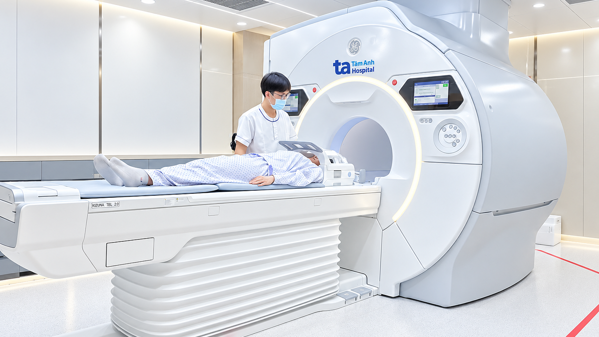
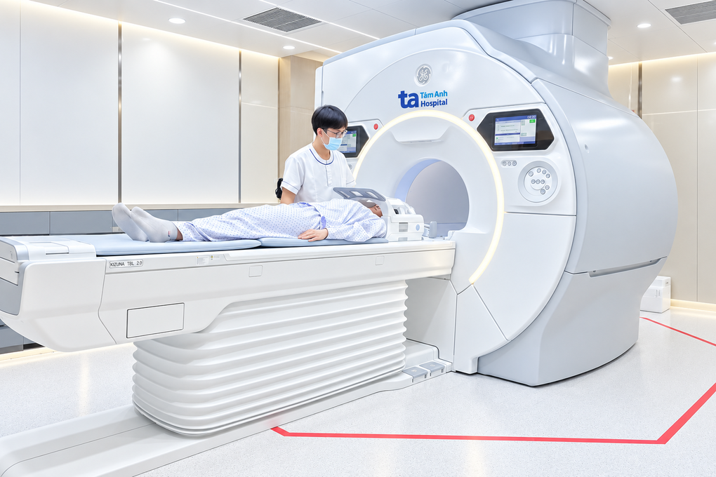
Describing the technology, the doctor gave the example of the case of Khang, 32 years old. The patient was admitted to the hospital because of increasing headaches, weakness in his right hand, and many times he dropped things. A 3 Tesla MRI scan showed that Mr. Khang had a tumor about 4.5 cm in the left frontal lobe, infiltrating surrounding brain tissue, near the motor cortex and nerve fiber bundles that control movement. Due to the infiltrative nature, the boundary between tumor tissue and healthy tissue is difficult to recognize.
According to Dr. Tan Si, the surgeon needs to remove as much of the tumor as possible to avoid the remaining tumor from continuing to grow. At the same time, it is necessary to avoid cutting too widely, causing damage to healthy brain tissue, leading to the patient becoming weak, paralyzed or having other long-term neurological function defects. Meanwhile, the boundary between suspected tumor tissue and surrounding healthy tissue is not always clear.
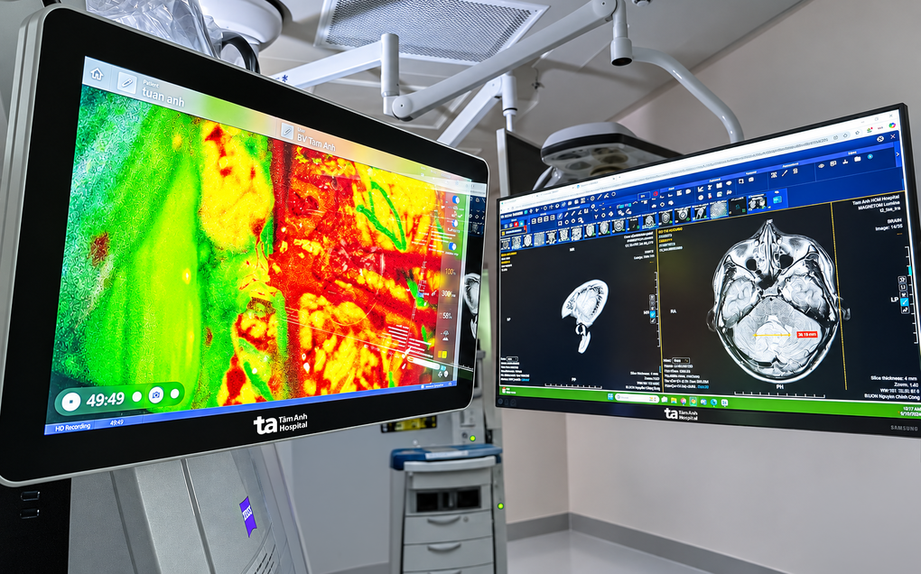
Therefore, the surgical team aims to remove maximum tumor tissue within safe limits, while preserving the patient's neurological function. They use luminescent tumor tissue dyeing technology. The doctor injects fluorescein sodium intravenously at a dose of 5 mg/kg, stimulates it with a suitable light source so that this substance emits fluorescent light and observes the signal with a new generation microsurgery integrated with artificial intelligence (AI) Zeiss Kinevo 900 with Yellow 560 mode. This is a fluorescence observation mode integrated in the microsurgery, highlighting areas of fluorescein accumulation.
After about 60-90 minutes, the doctor can observe the color, structure and morphology of brain tissue, and have additional visual signs to identify tumor edges (tumor boundaries), especially transition areas that are difficult to distinguish.
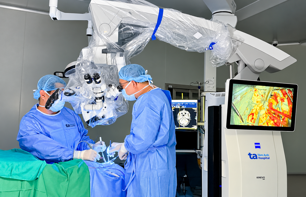
Dr. Tan Si explained that the surgical procedure is that the doctor will first remove the majority of the tumor under white light and then switch the microsurgical microscope to Yellow 560 mode for examination. The doctor compares these points with DTI diffusion tensor magnetic resonance imaging, nerve fiber maps and positioning systems to observe and evaluate suspected tumor tissue before deciding whether to remove any more tumor tissue.
If the tumor tissue location still has signal, it is a safe distance from the motor brain area and the corticospinal tract, the team will take more tumor tissue and re-examine the surgical field. If the signal appears too close to healthy brain structures, the doctor will stop expanding the cutting area.
Mr. Khang's surgery lasted nearly three hours, about 97.2% of the tumor volume was removed. Khang was alert, no further complications were noted. After three days, he had less headache and could sit up and walk around the room.
After surgery, the doctor continues to evaluate the molecular characteristics of the tumor to develop a treatment regimen. Patients can receive radiotherapy combined with chemotherapy to control remaining tumor cells, reducing the risk of disease progression or recurrence.
This fluorescent tumor tissue dyeing technology has just been reported by Tam Anh General Hospital at the hospital's 2026 International Scientific Conference. Accordingly, 35 high-grade astrocytoma patients have had this technology applied for surgery, the surgeries took place from September 2024 to September 2026. On average, 95.8% of the tumor volume seen on MRI was removed. In the group with clear fluorescent signals, the average rate reached 97.6%.
Evaluating the technology, Dr. Tan Si said the biggest benefit is that the surgeon will have more accurate grounds to distinguish the suspected tumor area from the surrounding healthy brain tissue, especially when the tumor boundary is unclear under white light.
This technology is especially effective in patients with brain tumors, such as high-grade astrocytoma. This is a malignant brain tumor that can grow quickly and spread into surrounding brain tissue. Tumor surgery plays a key role in treatment, with the goal of removing the maximum amount of diseased tissue possible, then combining additional treatments such as radiotherapy and chemotherapy depending on the case. The higher the tumor removal level, the less remaining diseased tissue, thereby making it more convenient for disease control and post-operative treatment.
"Removing more malignant tumor tissue within safe limits helps reduce the amount of remaining tumor cells, facilitates subsequent radiotherapy and chemotherapy, and contributes to the decision to prolong disease control time," the doctor said.
For patients, when the maximum tumor portion is removed within safe limits, while healthy brain areas are protected, they have more opportunities to maintain movement, language, cognition and the ability to exercise autonomy in daily life. This is an important goal to improve prognosis and quality of life, rather than just focusing on getting through surgery.
In the coming time, Tam Anh General Hospital in Ho Chi Minh City plans to expand technical applications for suitable brain tumor cases, while combining fluorescence imaging with nerve localization, imaging of nerve fiber bundles and a system for monitoring nerve function during surgery; Standardize the process of using fluorescein to stain tumor tissue, build case data and train to expand the surgical team, better serving brain tumor patients, which is a dangerous disease.
Van Ha